
The outer ear includes the portion of the ear that we see—the pinna/auricle and the ear canal.
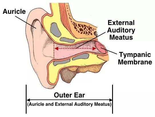
The ear canal or external auditory meatus is approximately 1.25 inches long and .25 inch in diameter. The inner two-thirds of the ear canal is imbedded in the temporal bone. The outer one-third of the canal is cartilage. Although the shape of each ear canal varies, in general the canal forms an elongated “s” shape curve. The ear canal directs airborne sound waves towards the tympanic membrane (eardrum). The ear canal resonates sound waves and increases the loudness of the tones in the 3000-4000 Hz range.
The ear canal maintains the proper conditions of temperature and humidity necessary to preserve the elasticity of the tympanic membrane. Glands, which produce cerumen (earwax) and tiny hairs in the ear canal, provide added protection against insects and foreign particles from damaging the tympanic membrane.
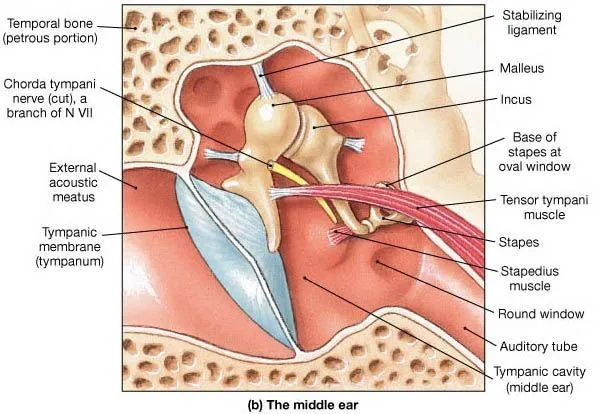
The middle ear is composed of the tympanic membrane and the cavity, which houses the ossicular chain.
The tympanic membrane or eardrum serves as a divider between the outer ear and the middle ear structures. It is gray-pink in color when healthy and consists of three very thin layers of living tissue.
The eardrum is very sensitive to sound waves and vibrates back and forth as the sound waves strike it. The eardrum transmits the airborne vibrations from the outer to the middle ear and also assists in the protection of the delicate structures of the middle ear cavity and inner ear.
The middle ear is connected and transmits sound to the inner ear via the ossicular chain. The ossicular chain amplifies a signal approximately 25 decibels as it transfers signals from the tympanic membrane to the inner ear.
The ossicular chain consists of the three smallest bones in the body: the malleus, incus, and stapes. The malleus is attached to the tympanic membrane. The footplate of the stapes inserts into the oval window of the inner ear. The incus is between the malleus and the stapes.
Attached to the ossicular chain are two tiny muscles, the stapedius and tensor tympani muscles. These muscles contract to protect the inner ear by reducing the intensity of sound transmission to the inner ear from external sounds and vocal transmission.
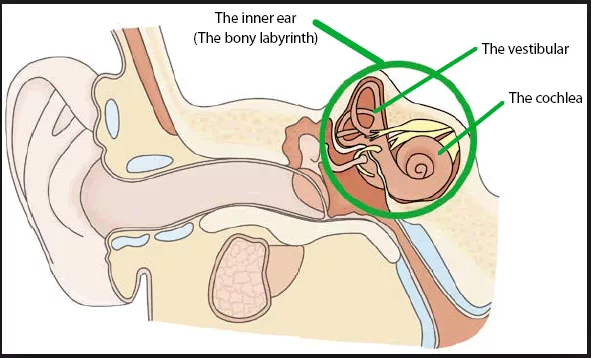
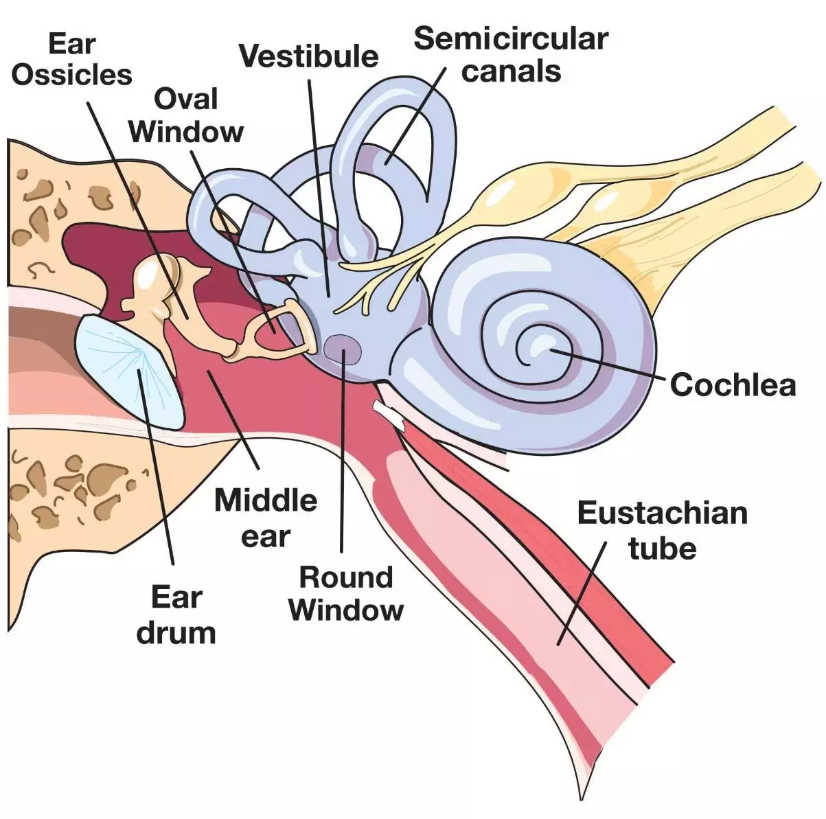
The inner ear is composed of the sensory organ for hearing—the cochlea, as well as for balance—the vestibular system. The systems are separate, yet both are encased in the same bony capsule and share the same fluid systems.
The cochlea is composed of three fluid-filled chambers that extend the length of the structure. The two outer chambers are filled with a fluid called perilymph. Perilymph acts as a cushioning agent for the delicate structures that occupy the center chamber. It is important to note that perilymph is connected to the cerebrospinal fluid that surrounds the brain and the spinal column. The third fluid filled chamber is the center chamber, called the cochlear duct. The cochlear duct secretes a fluid called endolymph, which fills this chamber.
The cochlear duct contains the Basilar membrane upon which lies the Organ of Corti. The Organ of Corti is a sensory organ essential to hearing. It consists of approximately 30,000 finger-like projections of cilia that are arranged in rows. These cilia are referred to as hair cells. Each hair cell is connected to a nerve fiber that relays various impulses to the cochlear branch of the VIIIth cranial nerve or auditory nerve. The “pitch” of the impulse relayed is dependent upon which areas of the basilar membrane, and hence, which portions of the Organ of Corti are stimulated. The apical portion of the basilar membrane (the most curled area of the cochlea) transfers lower frequency impulses. The basal end relays higher frequency impulses.
The VIII cranial nerve (VIII C.N.) or auditory C.N. carries the impulses generated from the Organ of Corti to the brainstem. From the brainstem, nerve pathways extend through numerous nuclei to the cerebral cortex in the temporal lobes of the brain. It is in the temporal lobes of the brain that meaning is associated with the various patterns of nerve impulses.
The Path Of Sound Until Perception
Sound Transmission through the Outer Ear
Air transmitted sound waves are directed toward the delicate hearing mechanisms with the help of the outer ear, first by the pinna, which gently funnels sound waves into the ear canal, then by the ear canal.
Sound Transmission through the Middle Ear
When air movement strikes the tympanic membrane, the tympanic membrane or eardrum moves. At this point, the energy generated through a sound wave is transferred from a medium of air to that which is solid in the middle ear. The ossicular chain of the middle ear connects to the eardrum via the malleus, so that any motion of the eardrum sets the three little bones of the ossicular chain into motion
Sound Transmission through the Inner Ear
The ossicular chain transfers energy from a solid medium to the fluid medium of the inner ear via the stapes. The stapes is attached to the oval window. Movement of the oval window creates motion in the cochlear fluid and along the Basilar membrane. Motion along the basilar membrane excites frequency specific areas of the Organ of Corti, which in turn stimulates a series of nerve endings.
Sound Transmission to the Brain
With the initiation of the nerve impulses, another change in medium occurs: from fluid to neural. Nerve impulses are relayed through the VIII C.N., through various nuclei along the auditory pathway to areas to the brain. It is the brain that interprets the neural impulses and creates a thought, picture, or other recognized symbol.
Appointments
All patients are seen on an appointment basis. Should you wish to make a booking, please contact our receptionists on
(011) 482-5530 and they will assist you in finalising a suitable date and time,
Alternatively, click on the button below.